

January 07, 2026
Goodbye Painful IVF Hormone Injections
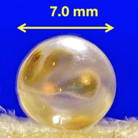
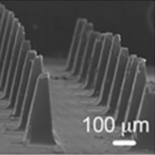
Our work on a light-triggered microneedle patch for painless, automated IVF hormone delivery was featured in the OMNI Television, CBC Listen, CTV News, LA PRESS, and McGill Newsroom.
At the Biointerface Lab we strive to understand and control
phenomena occurring at the interface between synthetic
materials and biological molecules. We have three main areas
of focus: biomineralization, i.e. the formation of minerals
in living organisms, both physiological and pathological,
implant-tissue integration, and drug delivery. An emerging
aspect of our research is the exploration of the
intersection between science and art. Explore more details
below!
Students trained in our group gain a multidisciplinary
skillset in chemistry, biology, and materials science and
engineering. Past lab members
are now successfully employed in academia or industry, or
pursue advanced degrees.


Click on the images to learn more or view a full list of
publications from our group!
We are excited about sharing our research with the general public!
For a full timeline of media coverage of our research click here!
Our work on a light-triggered microneedle patch for painless, automated IVF hormone delivery was featured in the OMNI Television, CBC Listen, CTV News, LA PRESS, and McGill Newsroom.
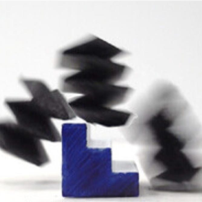
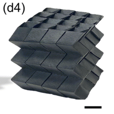
Our work on magnetoactive graphene oxide sheets that rapidly fold into reprogrammable soft robots and load-bearing origami structures was featured as a Nanowerk spotlight.
She was awarded the CRC to study Sex, Age and
Calcification Diseases. Her appointment was covered in
two press releases in the
McGill Reporter and on the
McGill website. Congratulations Marta!
The biointerface lab is equipped with state-of-the-art
spectrometers, sepcifically designed for the analysis of
sample surfaces and interfaces. We also have a setup for the
synthesis of nano-microparticles in highly controlled
conditions. If you are interested in acessing any of these
instruments, please contact us.

We have a fantastic Bruker Senterra confocal Raman
microscope equipped with 3 lasers (785, 633 and 532 nm),
which allows us to analyze a vast range of samples,
including biological samples. A special hardware feature
removes fluorescence background at 785 nm. Sample maps with
~1 micron resolution can be obtained with a computer
controlled sample stage, and polarizers and analyzers allow
us to take polarized spectra. Additionally, we can measure
fluorescence maps using the Olympus optical microscope
installed on the instrument. The connection with the
MultiRAM allows us to measure spectra with excitation
wavelength of 1064 nm, too. This instrument is now
managed by MIAM. Please contact Lihong
Shang if you would like to access it.
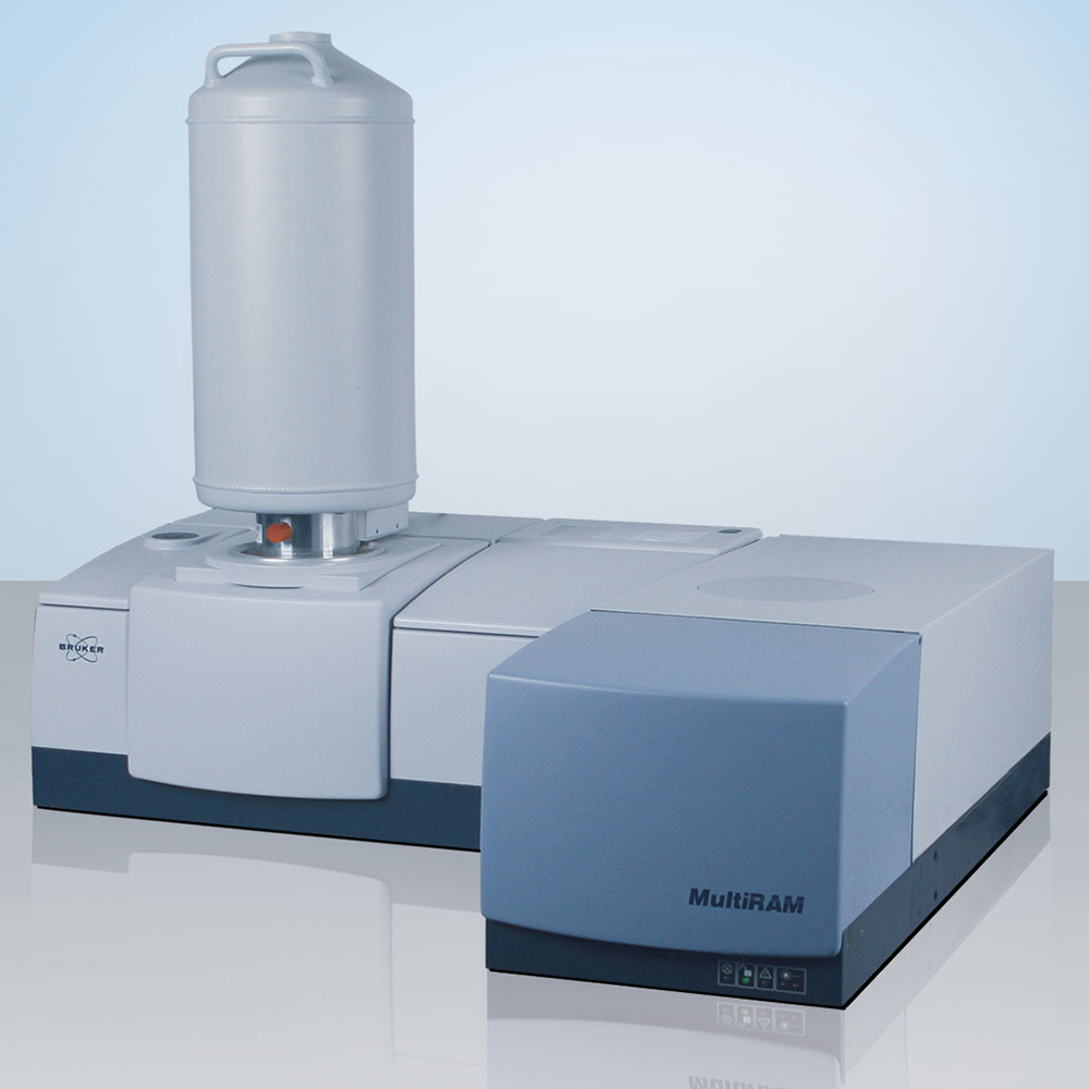
The Bruker MultiRAM spectrometer is a stand-alone FT-Raman spectrometer with excitation wavelength of 1064 nm. We have connected this instrument to the Senterra microscope via a fiber optic, thus achieving the rare possibility of collecting FT-Raman spectra inside a confocal microscope.
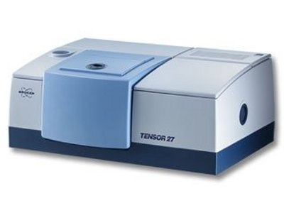
We have a Bruker Tensor 27 IR spectrometer, equipped with both a DTGS and an MCT detector. Several attachments allow us to perform measures specifically at interfaces: multi-pass attenuated total reflectance (ATR), with which we can take spectra of samples in water; diamond single-pass ATR, with which we can analyze surface layers; grazing-angle ATR (GATR), the best technique to analyze self-assembled monolayers; and a diffuse reflectance (DRIFT) cell.
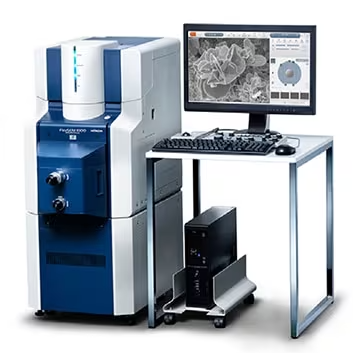
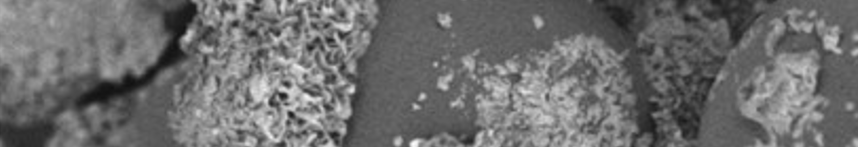
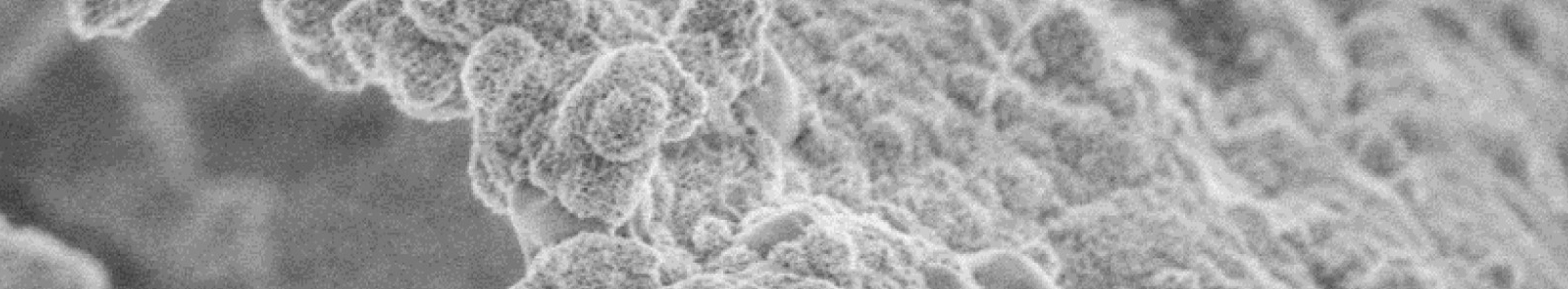
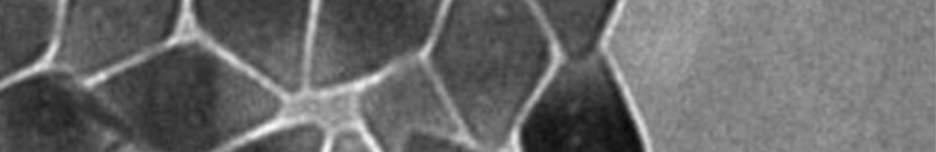
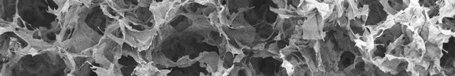
The tabletop FlexSEM 1000 SEM (Hitachi) allows us to take high resolution SEM images in secondary and backscattered electron modes, both in high vacuum or variable pressure mode. An EDS detector allows us to also measure surface composition of the materials we analyze.